

Myelogram
A myelogram is a diagnostic test performed to assess for compression of a spinal nerves or the spinal cord in either the lumbar (lower spine) or cervical spine (upper spine or neck) respectively. Commonly, this information can be gleaned using MRI but on occasion, it is not possible to undergo an MRI (for example if you have a pacemaker) or sometimes, information on neural compression in different physical positions (dynamic compression) is required to guide treatment. In these situations, a myelogram may be a sensible investigation to perform.
What happens before the procedure?
Before being referred for an myelogram, you need to be assessed by a specialist surgeon or physiotherapist who will take a history of your symptoms and perform a thorough examination. If indicated, an MRI scan or CT scan may be requested to investigate your symptoms. These tests will help to locate which level in the spine your pain or other neurological symptoms such a numbness may be emanating from and which nerves are most likely to be the cause of the symptoms. If it is decided that you may benefit from further investigation with a myelogram, we assess your prior imaging to guide the examination.
How do I prepare for my procedure?
Please inform us if you take regular blood thinning medication when you arrange your appointment as some need to be temporarily withheld beforehand. Do not stop any medication before talking to us. This may be an antiplatelet medication (such as aspirin or clopidogrel), or an anticoagulant medication (for example warfarin, apixaban, dabigatran, or rivaroxaban)
How is the procedure performed?
The test involves placement of a needle into the spinal canal (a fluid filled cavity in the spine containing the spinal cord and nerve roots) and injection of X-ray contrast dye which dissolves into the spinal fluid to fill the space and allow identification of areas of external compression (usually from abnormal discs or vertebral bones which have undergone wear-and tear).
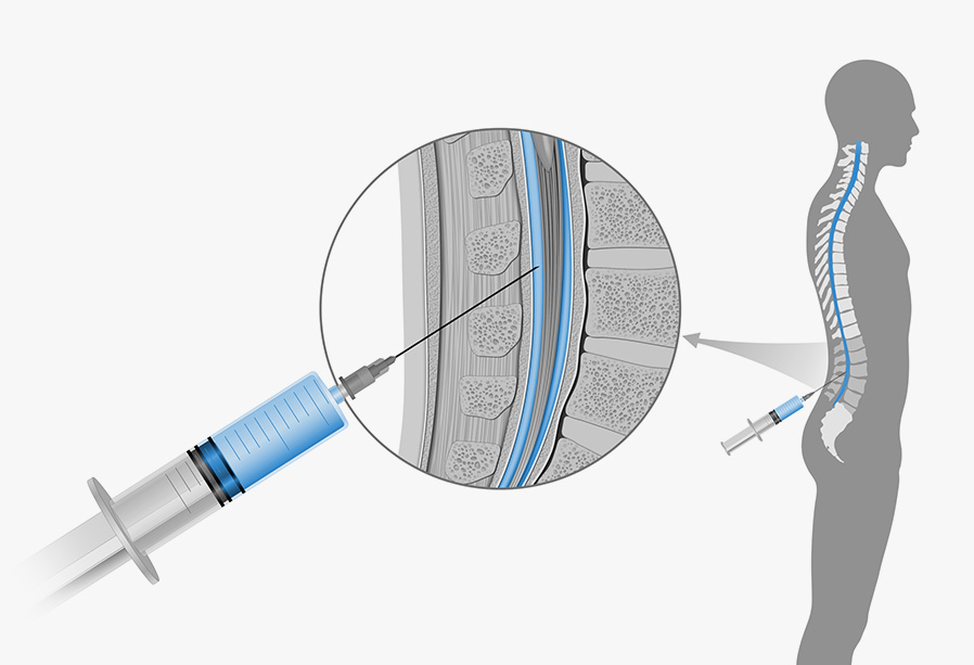
The procedure is commonly performed with the patient on their front. The back is cleaned and local anaesthetic injected into the skin to numb it. A thin spinal needle is the inserted under X-ray guidance to the appropriate position which is confirmed with a small volume of X-ray contrast dye. A larger volume of X-ray dye is then injected under X-ray surveillance and the needle is then removed.
We then perform a number of manoeuvres including tilting the X-ray table or asking the patient to role in-order to mix the dye within the spinal fluid. If we are aiming to image the neck, we often require quite significant tilting angles in order that the dye can travel to the neck with gravity. In these situations we commonly place the patient in safety straps.
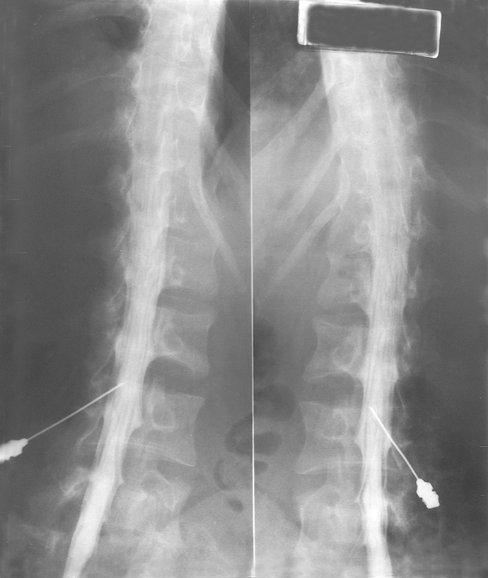
A number of X-rays are then performed. This may be with the patient in a standing, leaning or arched position to image the lower back.
If we are aiming to image the neck or cervical spine, we will image the neck in flexed or extended positions. We usually then perform a CT of the target area to get more information. This is performed in a separate room shortly after the injection of dye.
What are the side effects of the procedure?
The most common side effect of the injections are slight tenderness and/or bruising at the site of the injection, this usually resolves over the first few days. Patients can describe a leg cramping sensation after injection of the xray dye which will pass in a short time. Another potential side effect is of headache due to leakage of spinal fluid (fluid that bathes the brain and spinal cord) and subsequent reduction of the pressure of this fluid. This is most commonly self-limiting and can be treated using hydration, simple pain killers and increased caffeine intake. Very occasionally the procedure can be complicated by bleeding or infection.